What You Need to Know About Retinal Tears
Admin • May 1, 2020
The complex processes that enable vision involve some relatively delicate tissues, including the light-sensitive tissue at the back of the eye known as the retina. If this tissue becomes torn, you may experience a number of severe visual issues, potentially including an eventual retinal detachment and permanent vision loss.
You can protect your eyesight more effectively if you understand what kinds of threats your retinas face, what trouble signs to watch out for, and what kind of care to seek. Here are some essential points about retinal tears and related retinal damage.
How the Retina Functions
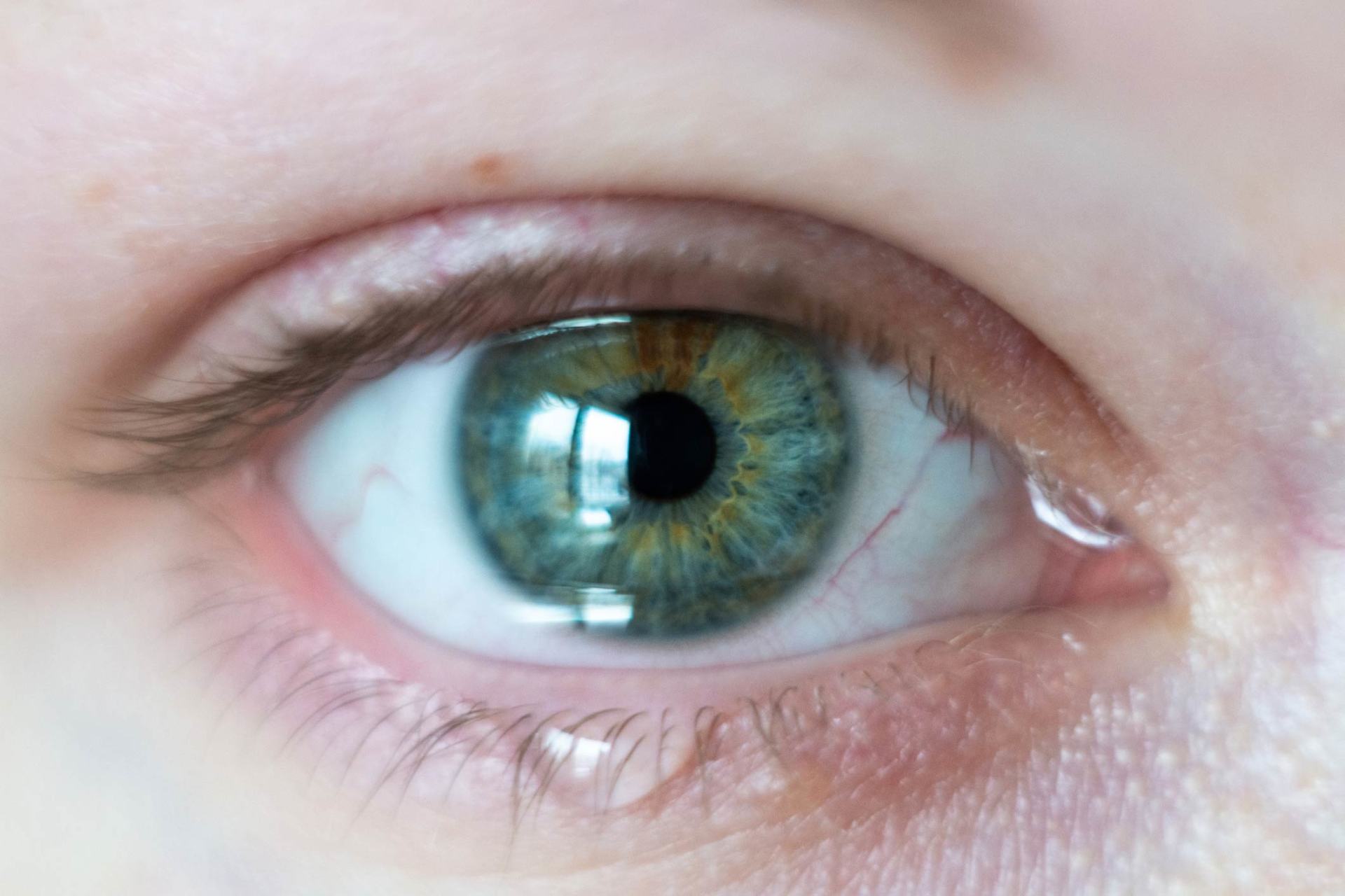
The multi-layered tissue of the retina lines most of the eye's interior surface. The retina contains numerous tiny nerve cells called rods and cones. When incoming light passes through the lens of the eye, it strikes the retina and causes these cells to transmit visual data to the optic nerve, which in turn relays them to the brain.
The cones tend to occupy a zone of the retina called the macula, conveying the data that provide crisp, clear central vision under bright conditions. The rods extend outward from this zone, receiving the signals that make up your peripheral and low-light vision.
How Retinal Tears Happen
A clear, gelatinous fluid known as the vitreous fills the inner globe of the eye and rests against the retinal wall. With age, however, this fluid tends to change in consistency, becoming more liquid in nature and hosting stringy filaments. These bits of debris create the spots or shapes many people perceive as floaters .
As the vitreous loses its gelatinous nature and shrinks, it pulls away from the retina. Occasionally this tugging action can cause a tear in the retinal wall. In some cases, blood vessels may also become torn, leaking into the eye or behind the retinal wall.
What Retinal Tears Do to Your Vision
When a retinal tear occurs, you may experience a sudden onset of floaters or flashes (sparkles of bright light). With luck, these symptoms may subside on their own over time as the retinal tear manages to heal itself.
A retinal tear that doesn't succeed in healing itself may grow progressively larger if untreated. This progress, coupled with ongoing bleeding behind the retina, can eventually cause part of the retinal wall to detach from the inner surface of the eye. A detached retina may appear as a dark curtain across your vision.
How Ophthalmologists Treat Retinal Tears
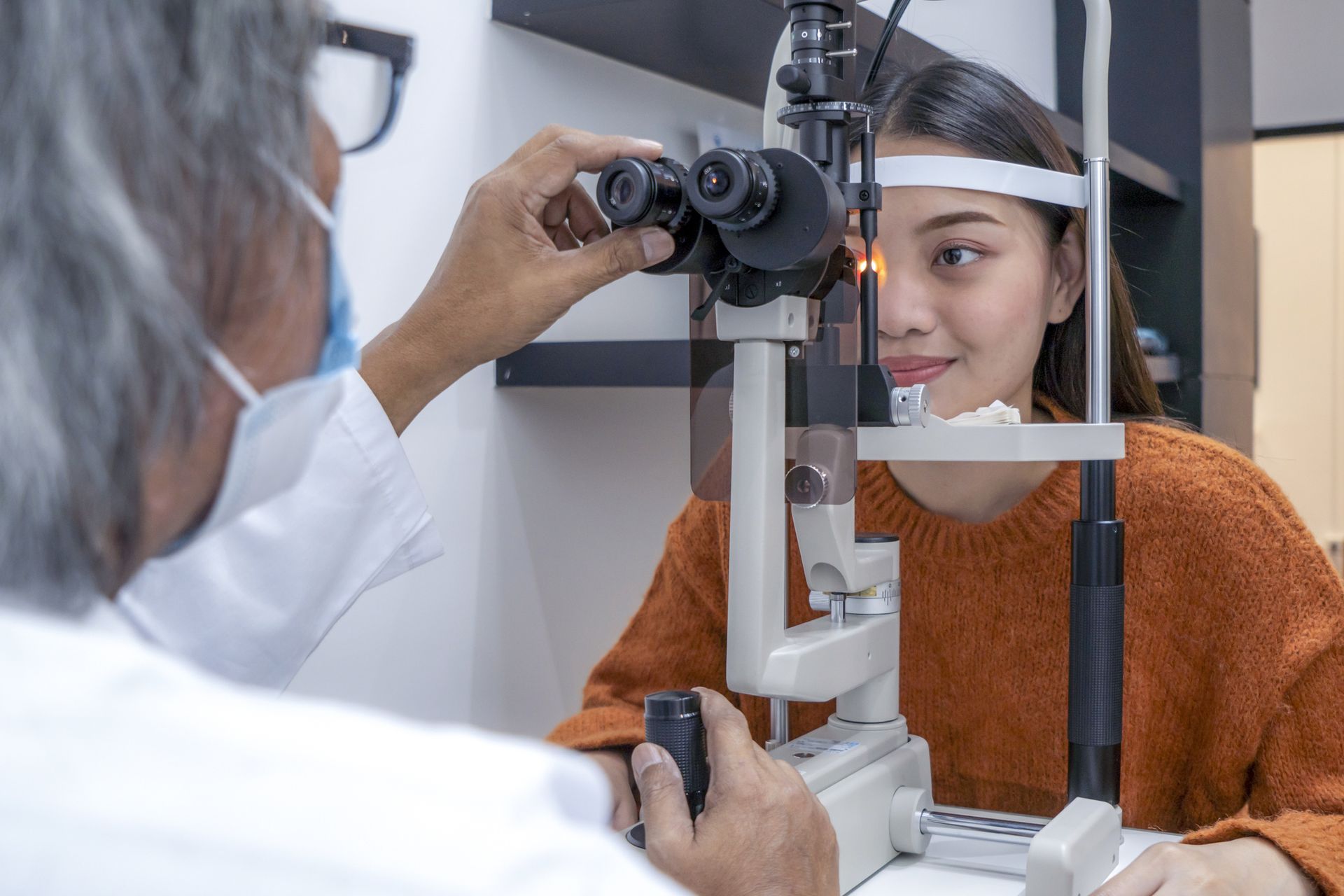
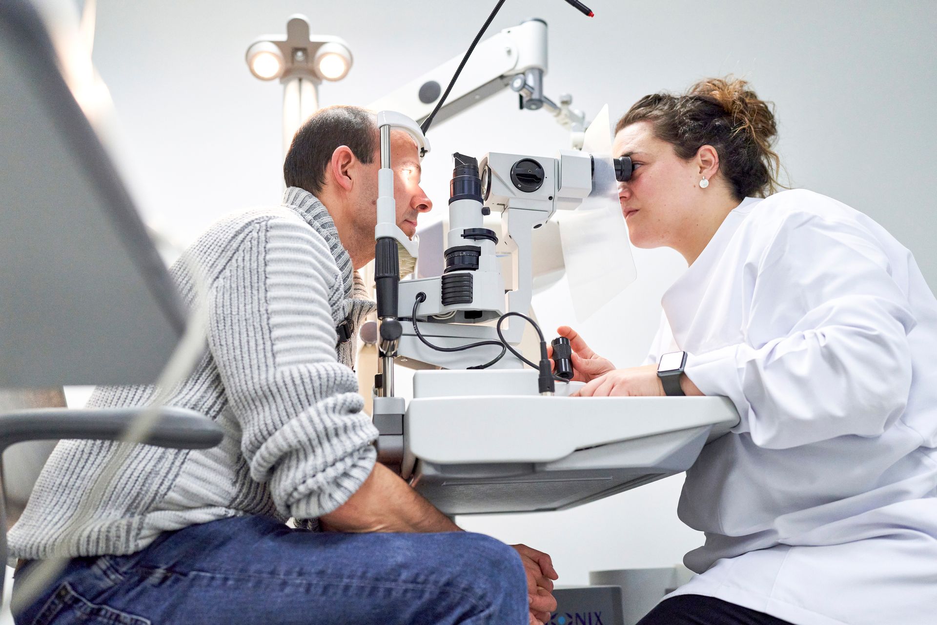
Age-related floaters and flashes represent a relatively common problem, especially in middle-aged or senior individuals. An ophthalmologist can inspect your retinas for any signs of tears. Small tears may require no treatment apart from ongoing monitoring through regular eye exams.
Retinal tears that appear to present a danger to your eyesight require immediate treatment to keep them from growing into full-scale detachments. Laser treatment can stop small tears by creating scar tissue on the tear's edges and preventing further damage or blood leakage. Extreme cold can accomplish similar results.
Your ophthalmologist may also decide that you need for fluid in your eye to counteract shrinking of the vitreous and prevent future tears. An injection of clear silicon can take the place of your natural vitreous if necessary.
If your retinal tear progresses to the detachment stage, you may need more invasive surgery to repair this kind of damage and remove the fluid trapped behind the retinal wall. Fortunately, ophthalmologists can usually successfully reattach detached retinas with timely diagnosis and treatment.
If you suspect that you may have a retinal tear, Calvert Ophthalmology Center can evaluate your eyes and recommend the proper course of treatment. Contact one of our ophthalmology offices today for an appointment.

Find out when to visit a vision care doctor in Clarksville, TN, at Calvert Ophthalmology Center. Call (931) 552-2233 for expert eye health care and advice.
Get thorough eye exams in Nashville, TN, at Calvert Ophthalmology Center. Schedule your appointment today for expert, personalized eye care.

Discover 5 key benefits of expert optometry in Franklin, TN. Protect your vision with care from Calvert Ophthalmology Center. Contact us at (931) 552-2233.
Discover 5 key tips for preparing for eye exams in Nashville, TN. Schedule your appointment with Calvert Ophthalmology Center today!

Learn how to maintain good eye health with tips from optometry experts in Franklin, TN. Call Calvert Ophthalmology Center today for professional assistance.
Glasses and contacts are two of the most popular choices for improving vision. For assistance deciding which is right for you, read this guide.
Despite diligent care for the muscles and bones, it is unfortunate that athletes often overlook eye health. Read this blog to learn more.
Vision problems can be common as people age. If you're getting older and want to learn more, check out our blog to read about a few common eye problems.
Pink eye can affect both kids and adults. Learn more with this overview of the causes, symptoms, and options for treatment of this eye condition.
Do you wear contact lenses? If so, read our blog to learn about the telltale signs that indicate it's time to replace your lenses.